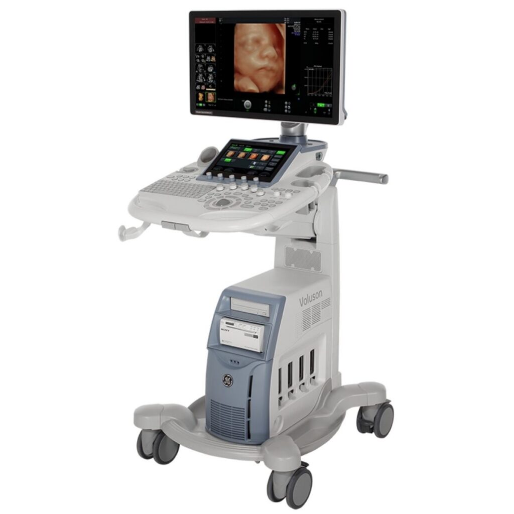
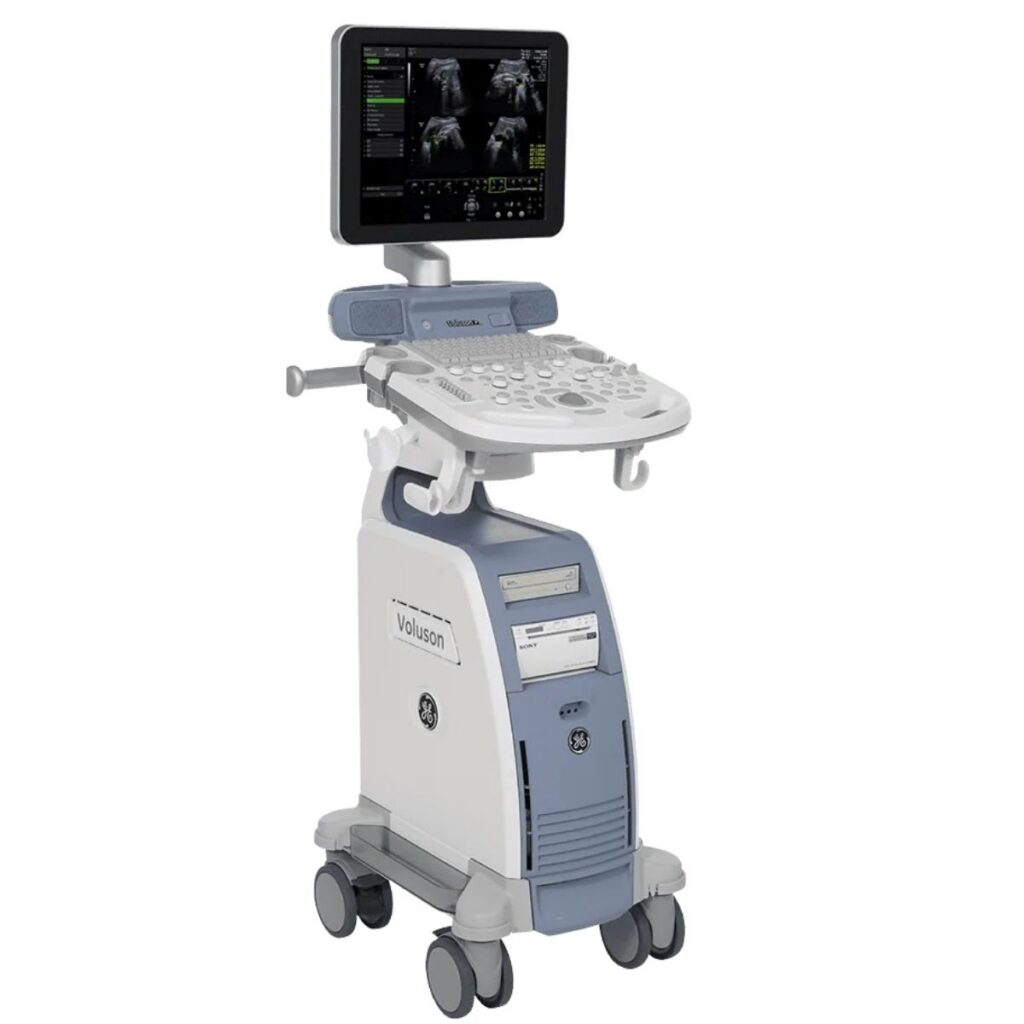
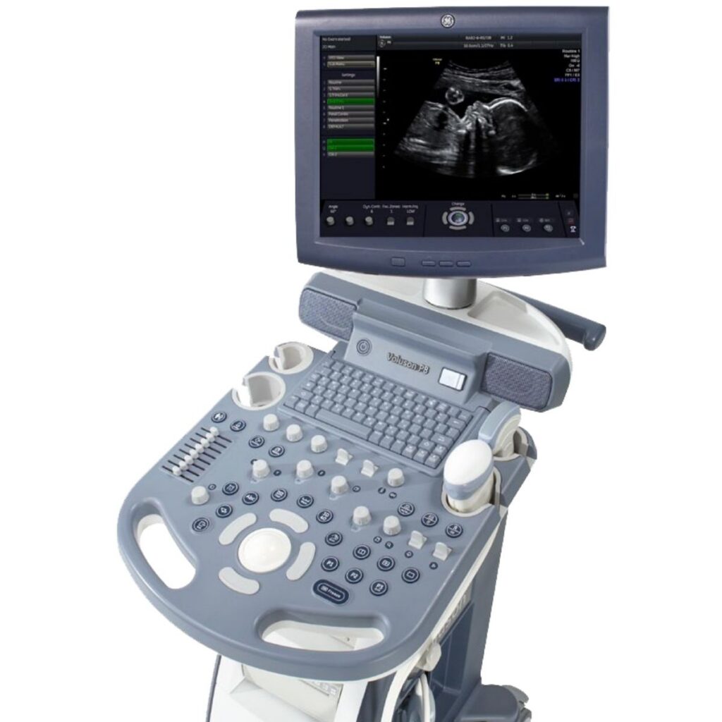
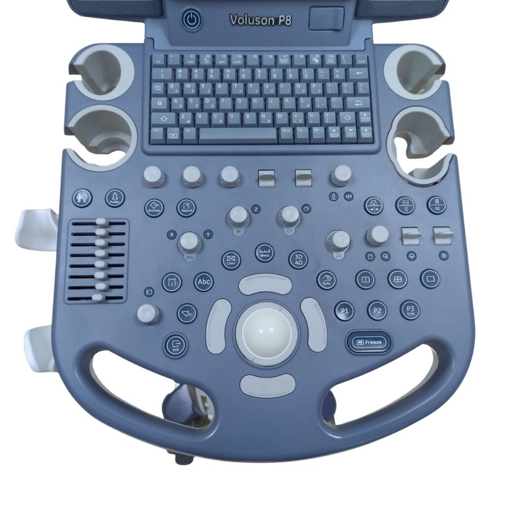
GE VOLUSON P8

Versatile refurbished ultrasound system designed for obstetric and gynecological imaging, combining high-quality 2D, Doppler and 3D/4D imaging with an intuitive workflow.

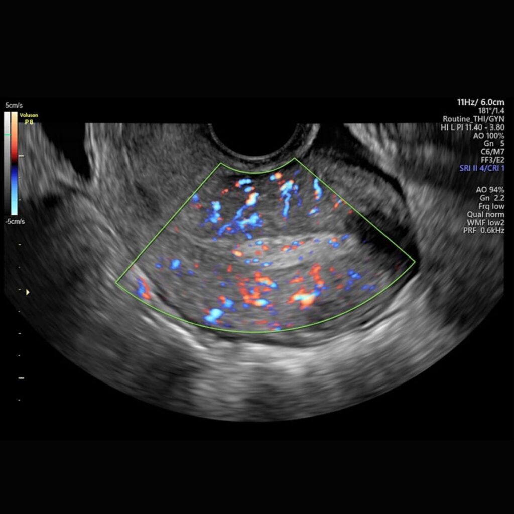
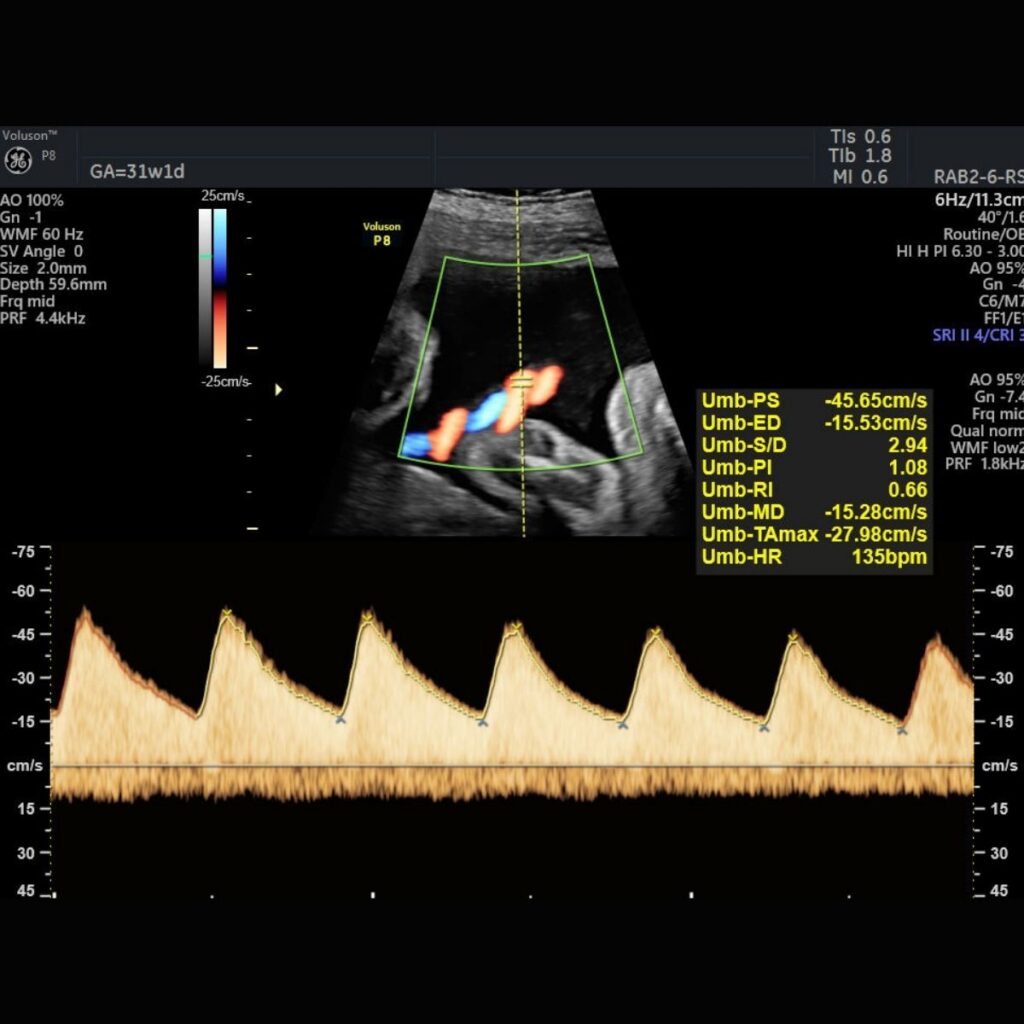
The GE Voluson P8 is a versatile ultrasound system developed for obstetric and gynecological imaging. It combines reliable 2D image quality, Color and Pulsed Wave Doppler, 3D/4D volume imaging and workflow automation tools in a compact and intuitive platform. Designed to support routine examinations as well as more advanced women’s health applications, the Voluson P8 helps healthcare professionals perform efficient examinations while maintaining consistent image quality and streamlined reporting.

ABOUT THIS SYSTEM

Reliable Imaging for Women’s Health


The GE Voluson P8 brings established Voluson imaging technology into a compact platform designed for everyday women’s health applications. Its capabilities support obstetric evaluations, gynecological studies, fertility screening and embryo transfer procedures, while advanced imaging tools help improve visualization and diagnostic confidence.
The system is designed to simplify daily workflow through intuitive controls, automated optimization and structured reporting tools. 3D and 4D imaging provide additional anatomical perspectives and allow volume data to be reviewed during or after the examination.

TECHNICAL SPECIFICATIONS

  • 2D Imaging
    High-quality 2D imaging designed to provide clear visualization across routine obstetric and gynecological examinations.
  • Color and Pulsed Wave Doppler
    Doppler technologies support the evaluation of vascular anatomy and blood flow.
  • 3D/4D Imaging
    Volume imaging allows the acquisition, analysis and manipulation of volumetric data during the examination or offline, with multiple rendering methods available.
  • SRI – Speckle Reduction Imaging
    Helps improve visualization and contrast resolution by reducing speckle-related artifacts.
  • CrossXBeamCRI
    Helps enhance tissue interfaces and border differentiation.
  • HD-Flow
    Bi-directional Power Doppler technology designed to improve sensitivity in vascular studies.
  • SingleView
    Supports visualization of anatomy in coronal or semi-coronal planes with selectable slice thickness.
  • Intuitive workflow
    The control panel is designed to provide quick access to frequently used functions and support efficient examinations.
  • Automated image optimization
    Auto Optimization for 2D and Doppler helps improve image contrast and spectral Doppler settings at the touch of a button.
  • Auto Focus
    Automated focal-zone positioning helps reduce manual adjustments during 2D scanning.
  • Raw Data
    Images can be stored in their original quality, allowing post-exam review and additional image adjustments without repeating the examination.
  • Structured reporting
    Reporting tools support obstetric and gynecological examinations and allow reports, charts and measurements to be exported in PDF format.
  • Wireless connectivity
    Supports efficient transmission of images and reports.
  • Compact system design
    The Voluson P8 is designed with a relatively small physical footprint to support use in clinical environments where space efficiency is important.

Obstetrics

  • Routine obstetric examinations
  • First-trimester assessment
  • Second- and third-trimester examinations
  • Fetal anatomy evaluation
  • Fetal cardiac imaging
  • Doppler assessment
  • 3D/4D fetal imaging

Gynecology

  • Pelvic imaging
  • Uterine assessment
  • Ovarian evaluation
  • Gynecological volume imaging


Fertility and Reproductive Medicine

  • Fertility screening
  • Follicular assessment
  • Embryo transfer support

The GE Voluson P8 supports a range of abdominal and endocavitary transducers designed for obstetric and gynecological imaging. Available accessories and probes depend on the specific configuration of the system. Please contact SONOS for information about the accessories included with this unit.

Depending on system configuration, the GE Voluson P8 can support a range of advanced technologies designed to improve imaging quality and workflow:

  • 3D/4D volume imaging
  • SonoRender / SonoRenderlive
  • SonoBiometry
  • SonoNT
  • SonoIT
  • SRI – Speckle Reduction Imaging
  • CrossXBeamCRI
  • HD-Flow
  • SingleView
  • Auto Optimization for 2D and Doppler
  • Auto Focus
  • Wide-sector imaging
  • Raw Data
  • Structured reporting and PDF export
  • Wireless connectivity

Availability of individual software packages and options depends on the specific system configuration.

DOWNLOADS

GE Voluson P8 Brochure

Need more information about this ultrasound system?

Our specialists can provide technical details, availability and support to help you find the right ultrasound solution.